| Sel parafolikel | |
|---|---|
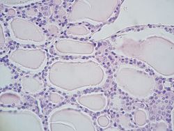
 Bahagian mikroskopik tiroid menunjukkan folikel yang dilapisi oleh sel epitel folikular, dan di antaranya sel parafollikular yang lebih besar. | |
| Butiran | |
| Lokasi | Tiroid |
| Fungsi | Merembeskan kalsitonin |
| Pengenalpastian | |
| TH | H3.08.02.4.00009 |
| FMA | FMA:68653 |
| Istilah anatomi mikroanatomi | |
Sel parafolikel, juga disebut sel C, adalah sel neuroendokrin di tiroid. Fungsi utama sel-sel ini adalah untuk mengeluarkan kalsitonin. Ia terletak bersebelahan dengan folikel tiroid dan berada di tisu penghubung. Sel-sel ini besar dan mempunyai noda pucat dibandingkan dengan sel folikel. Dalam spesies teleos dan burung, sel-sel ini menempati struktur di luar kelenjar tiroid yang dinamakan jasad ultimobrankium.
Struktur
Sel-sel parafolikular adalah sel-sel pewarnaan pucat yang terdapat dalam jumlah kecil pada tiroid dan biasanya terletak pada asasnya di epitel, tanpa hubungan langsung dengan lumen folikel. Selalunya terletak di dalam membran dasar, yang mengelilingi seluruh folikel.
Perkembangan
Sel parafolikular berasal dari endoderma farinks.[1][2] Secara embriologi, ia bersekutu dengan jasad ultimobrankium, yang merupakan turunan ventral dari kantung farinks keempat (atau kelima). Sel-sel parafolikular sebelumnya dipercayai berasal dari krista/rabung neural berdasarkan serangkaian eksperimen pada chimera burung puyuh.[3][4] Walau bagaimanapun, eksperimen penelusuran garis keturunan pada tikus menunjukkan bahawa sel parafollikular berasal dari asal endoderma. [5]
Fungsi
Sel parafolikular merembeskan kalsitonin, hormon yang mengambil bahagian dalam pengawalan metabolisme kalsium. Kalsitonin menurunkan kadar kalsium dalam darah dengan menghalang resorpsi tulang oleh osteoklas, dan rembesannya meningkat sebanding dengan kepekatan kalsium.[6]
Sel parafolikular juga diketahui mengeluarkan dalam jumlah yang lebih kecil beberapa peptida neuroendokrin seperti serotonin, somatostatin atau CGRP.[7][8][9] Ia juga mungkin berperanan dalam mengatur pengeluaran hormon tiroid secara lokal, kerana ia mengekspresikan hormon pelepasan tirotropin.[10][11]
Kepentingan klinikal
Apabila sel-sel parafolikular menjadi kanser, ia membawa kepada karsinoma medula tiroid.
Rujukan
- ^ "On the Origin of Cells and Derivation of Thyroid Cancer: C Cell Story Revisited". European Thyroid Journal. 5 (2): 79–93. July 2016. doi:10.1159/000447333. PMC 4949372. PMID 27493881.
- ^ Johansson, E., Andersson, L., Örnros, J., Carlsson, T., Ingeson-Carlsson, C., Liang, S., … Nilsson, M. (2015). Revising the embryonic origin of thyroid C cells in mice and humans. Development, 142(20), 3519–3528. http://doi.org/10.1242/dev.126581
- ^ "New studies on the neural crest origin of the avian ultimobranchial glandular cells--interspecific combinations and cytochemical characterization of C cells based on the uptake of biogenic amine precursors". Histochemistry. 38 (4): 297–305. March 1974. doi:10.1007/bf00496718. PMID 4135055.
- ^ "Thyrotropin induces the acidification of the secretory granules of parafollicular cells by increasing the chloride conductance of the granular membrane". The Journal of Cell Biology. 107 (6 Pt 1): 2137–47. December 1988. doi:10.1083/jcb.107.6.2137. PMC 2115661. PMID 2461947.
- ^ "Revising the embryonic origin of thyroid C cells in mice and humans". Development. 142 (20): 3519–28. October 2015. doi:10.1242/dev.126581. PMC 4631767. PMID 26395490.
- ^ Melmed S, Polonsky KS, Larsen PR, Kronenberg HM (2011). Williams Textbook of Endocrinology (ed. 12th). Saunders. m/s. 1250–1252. ISBN 978-1437703245.
- ^ "Ultrastructural localization of calcitonin, somatostatin and serotonin in parafollicular cells of rat thyroid". The Histochemical Journal. 16 (12): 1265–72. December 1984. doi:10.1007/bf01003725. PMID 6152264.
- ^ "Induction of a neural phenotype in a serotonergic endocrine cell derived from the neural crest". The Journal of Neuroscience. 7 (9): 2874–83. September 1987. doi:10.1523/JNEUROSCI.07-09-02874.1987. PMC 6569149. PMID 3305802.
- ^ "Separation of dissociated thyroid follicular and parafollicular cells: association of serotonin binding protein with parafollicular cells". The Journal of Cell Biology. 88 (3): 499–508. March 1981. doi:10.1083/jcb.88.3.499. PMC 2112761. PMID 7217200.
- ^ "Thyrotropin-releasing hormone gene expression in normal thyroid parafollicular cells". Molecular Endocrinology. 3 (12): 2101–9. December 1989. doi:10.1210/mend-3-12-2101. PMID 2516877.
- ^ "Functional expression of the thyrotropin receptor in C cells: new insights into their involvement in the hypothalamic-pituitary-thyroid axis". Journal of Anatomy. 215 (2): 150–8. August 2009. doi:10.1111/j.1469-7580.2009.01095.x. PMC 2740962. PMID 19493188.
Bacaan lanjut
- Kameda Y (October 1987). "Localization of immunoreactive calcitonin gene-related peptide in thyroid C cells from various mammalian species". The Anatomical Record. 219 (2): 204–12. doi:10.1002/ar.1092190214. PMID 3120623. S2CID 12517073.
- Kameda Y, Nishimaki T, Miura M, Jiang SX, Guillemot F (January 2007). "Mash1 regulates the development of C cells in mouse thyroid glands". Developmental Dynamics. 236 (1): 262–70. doi:10.1002/dvdy.21018. PMID 17103415. S2CID 24848963.
- Kameda Y, Nishimaki T, Chisaka O, Iseki S, Sucov HM (October 2007). "Expression of the epithelial marker E-cadherin by thyroid C cells and their precursors during murine development". The Journal of Histochemistry and Cytochemistry. 55 (10): 1075–88. doi:10.1369/jhc.7a7179.2007. PMID 17595340.
- Kameda Y, Ito M, Nishimaki T, Gotoh N (March 2009). "FRS2alpha is required for the separation, migration, and survival of pharyngeal-endoderm derived organs including thyroid, ultimobranchial body, parathyroid, and thymus". Developmental Dynamics. 238 (3): 503–13. doi:10.1002/dvdy.21867. PMID 19235715. S2CID 13504555.
- Kameda Y (March 2016). "Cellular and molecular events on the development of mammalian thyroid C cells". Developmental Dynamics. 245 (3): 323–41. doi:10.1002/dvdy.24377. PMID 26661795. S2CID 12161896.
- Baber EC (1876). "Contributions to the Minute Anatomy of the Thyroid Gland of the Dog". Philosophical Transactions of the Royal Society of London. 166: 557–568. doi:10.1098/rstl.1876.0021. JSTOR 109205.


